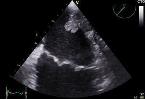
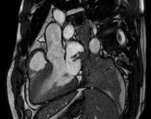
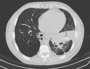
The patient was a 43-year-old man with a history of recurrent respiratory tract infection who attended our hospital with progressive dyspnea. Chest X-ray showed a left basal infiltrate and the electrocardiogram showed atrial fibrillation of unknown duration. Echocardiographic study showed a pedunculated mass by the lateral wall of the left atrium indicative of extracardiac origin (Figure 1 and Video 1 and Video 2 of the supplementary material). Cardiac magnetic resonance imaging and computed tomography confirmed atrial infiltration, arising from consolidation around a left hilar lymph node. This mass contained a foreign body measuring 22mm (Figure 2, Figure 3, and Video 3 of the supplementary material). Lung biopsy revealed abundant acute and chronic inflammatory infiltrate. In view of these findings, the patient was questioned again, and he recalled aspiration of a chicken bone a few years earlier. The patient was referred for left pneumonectomy and atrial surgery, although he later died of septic shock.


The clinical manifestations of bronchial aspiration usually occur immediately. However, there may be an asymptomatic interval if complete obstruction does not occur, especially with bones or inorganic material. In these cases, the process may resemble asthma or pneumonia due to inflammation and chronic superinfection. We believe that the delay in diagnosis of our patient enabled an inflammatory granuloma to develop around the foreign body, with infiltration of the left atrium due to its proximity.